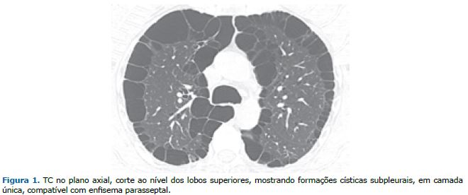
Homem, 35 anos, com queixas de tosse seca e dispneia progressiva há 2 anos, com piora recente. Usuário de maconha desde os 11 anos. A TC do tórax evidenciou múltiplas imagens císticas subpleurais, formando uma camada única (Figura 1).
Formações císticas subpleurais têm basicamente dois diagnósticos diferenciais: faveolamento e enfisema parasseptal. O faveolamento é um padrão tomográfico que consiste de múltiplos cistos agrupados em camadas e compartilhando paredes, em topografia subpleural. Histologicamente, faveolamento corresponde à formação de cistos pulmonares criados pela destruição de espaços aéreos distais, por fibrose do parênquima pulmonar, com desarranjo da arquitetura de ácinos e bronquíolos. Em síntese, a identificação de faveolamento pulmonar significa a presença de fibrose e é um importante critério para o diagnóstico de pneumonia intersticial usual.(1)
Enfisema é caracterizado patologicamente pelo alargamento anormal e permanente dos espaços aéreos distais ao bronquíolo terminal, com destruição das paredes alveolares. A classificação do enfisema é tradicionalmente baseada na localização da doença em relação ao lóbulo secundário. Os principais tipos são enfisema centroacinar ou centrolobular, parasseptal ou acinar distal e panacinar ou panlobular. Os achados tomográficos são de áreas de baixa atenuação, tipicamente sem paredes visíveis. No enfisema parasseptal ocorre um alargamento permanente da região distal do lóbulo pulmonar secundário, com destruição de ductos e sacos alveolares, com alvéolos dilatados em localização subpleural junto aos septos interlobulares. No enfisema parasseptal, as formas menos graves são de difícil detecção nas radiografias do tórax. Na TC são vistas formações císticas subpleurais eventualmente separadas por septos interlobulares intactos. Caracteristicamente, é delimitado pela superfície pleural ou septos interlobulares. As porções anteriores e posteriores dos lobos superiores e as porções posteriores dos lobos inferiores são mais frequentemente afetadas. Bolhas podem estar associadas a essa forma de enfisema. Se ocorrer isoladamente ou for o tipo predominante, o enfisema parasseptal tende a não causar quaisquer sintomas respiratórios e, portanto, muitas vezes é sub-reconhecido clinicamente.(2)
O enfisema parasseptal é mais comum em fumantes de maconha do que em fumantes apenas de tabaco. Parece que certas manobras realizadas pelos fumantes de maconha, como a inalação com manobra de Valsalva, podem levar a barotrauma e formação de bolhas apicais. O enfisema parasseptal é o padrão predominante observado em fumantes de maconha, enquanto o enfisema centrolobular é mais observado em fumantes apenas de tabaco. Isso pode representar um estágio anterior de formação de bolhas apicais relatadas em fumantes de maconha. Nosso paciente era um fumante pesado de maconha, que desenvolveu enfisema parasseptal.(3)
REFERÊNCIAS 1. Marchiori E, Hochhegger B, Zanetti G. Honeycombing. https://doi.org/10.1590/S1806-37562017000000232
2. Hochhegger B, Marchiori E, Rodrigues R, Mançano A, Jasinowodolinski D, Chate RC, et al. Consensus statement on thoracic radiology terminology in Portuguese used in Brazil and in Portugal [published correction appears in J Bras Pneumol. 2022 Jan 10;47(6):e20200595errata]. J Bras Pneumol. 2021;47(5):e20200595. https://doi.org/10.36416/1806-3756/e20200595
3. Murtha L, Sathiadoss P, Salameh JP, Mcinnes MDF, Revah G. Chest CT Findings in Ma-rijuana Smokers. Radiology. 2023;307(1):e212611. https://doi.org/10.1148/radiol.212611


 Read in English
 Portuguese PDF
 Print
Print
 Send this article by email
 How to cite this article
 Submit a comment
 Mendeley
 Pocket