ABSTRACT
Objective: To describe HRCT findings in patients with bacterial pneumonia following bone marrow transplantation (BMT). Methods: This was a retrospective study involving 30 patients diagnosed with bacterial pneumonia in whom HRCT of the chest was performed within 24 h after the onset of symptoms and the diagnosis was confirmed, based on a positive culture of sputum or bronchial aspirate, together with a positive pleural fluid or blood culture, within one week after symptom onset. There were 20 male patients and 10 female patients. The median age was 21 years (range, 1-41 years). The BMT had been performed for the treatment of the following: chronic myeloid leukemia, in 14 cases; severe aplastic anemia, in 6; acute myeloid leukemia, in 4; Fanconi's anemia, in 3; and acute lymphocytic leukemia, in 3. Two radiologists analyzed the HRCT scans and reached their final decisions by consensus. Results: The most common HRCT findings were air-space consolidation (in 60%), small centrilobular nodules (in 50%), ground-glass opacities (in 40%), bronchial wall thickening (in 20%), large nodules (in 20%), pleural lesions (in 16.7%) and tree-in-bud opacities (in 10%). The pulmonary lesions were distributed in the central and peripheral areas in 15 patients, whereas they were exclusively peripheral in 11. Lesions were located in the lower and middle lobes of the lung in 22 and 20 patients, respectively. Conclusions: The most common HRCT findings in our patient sample were air-space consolidation, small centrilobular nodules and ground-glass opacities, most often in the central and peripheral regions of the middle and lower lung zones.
Keywords:
Bone marrow transplantation; Pneumonia, bacterial; Tomography, X-Ray Computed.
RESUMO
Objetivo: Descrever os achados de TCAR em pacientes com pneumonia bacteriana após transplante de medula óssea (TMO). Métodos: Estudo retrospectivo com 30 pacientes diagnosticados com pneumonia bacteriana, documentada com TCAR do tórax realizada em até 24 h do início dos sintomas, e com diagnóstico comprovado com base em cultura positiva de escarro ou de aspirado brônquico associada à cultura positiva de líquido pleural ou de sangue dentro de uma semana após o início dos sintomas. Foram avaliados 20 pacientes masculinos e 10 femininos, com mediana de idade de 21 anos (variação, 1-41 anos). O TMO foi realizado para o tratamento de leucemia mieloide crônica (n = 14), anemia aplástica severa (n = 6), leucemia mieloide aguda (n = 4), anemia de Fanconi (n = 3) e leucemia linfoide aguda (n = 3). Dois radiologistas analisaram os exames de TCAR, chegando a decisões finais por consenso. Resultados: Os achados de TCAR mais frequentes foram consolidação do espaço aéreo (60%), pequenos nódulos centrolobulares (50%), opacidade em vidro fosco (40%), espessamento de parede brônquica (20%), nódulos grandes (20%), lesões pleurais (16,7%) e opacidades em padrão de árvore em brotamento (10%). As alterações pulmonares estavam distribuídas nas regiões centrais e periféricas dos pulmões em 15 pacientes e somente na periferia em 11 pacientes. As lesões estavam localizadas no terço inferior e no terço médio dos pulmões em 22 e 20 pacientes, respectivamente. Conclusões: Os achados de TCAR mais comuns na nossa amostra foram consolidações do espaço aéreo, pequenos nódulos centrolobulares e opacidades em vidro-fosco, distribuídos nas regiões centrais e periféricas dos terços médio e inferior dos pulmões.
Palavras-chave:
Transplante de medula óssea; Pneumonia bacteriana; Tomografia computadorizada por raios X.
IntroduçãoAs infecções pulmonares após transplante de medula óssea (TMO) são uma causa comum de morbidade e mortalidade.(1,2) Os patógenos mais comuns são as bactérias, fungos e vírus.(3-6) Poucos estudos anteriores discutiram os achados de TCAR em pacientes com pneumonia bacteriana após TMO.(4-14) O objetivo deste estudo foi descrever achados de TCAR em tais pacientes.
MétodosTrata-se de um estudo retrospectivo envolvendo 30 pacientes com pneumonia bacteriana, diagnosticados após TMO e confirmados através de estudos microbiológicos. Todos os pacientes assinaram um termo de consentimento livre e esclarecido, e a junta institucional de revisão do nosso hospital aprovou o estudo. Os pacientes haviam sido submetidos a TCAR, realizada em até 24 h do início dos sintomas da infecção, e um diagnóstico definitivo havia sido feito dentro de uma semana após o início dos sintomas. Naquela época, nenhum dos pacientes apresentou evidência de outra complicação pulmonar superposta. Os casos foram selecionados a partir de um grupo de pacientes que haviam sido submetidos ao TMO entre 1993 e 2006. Os casos com diagnóstico sem confirmação bacteriológica foram excluídos da análise. Em seguida, revisamos os registros médicos dos pacientes selecionados.
Dos 30 pacientes avaliados, 20 (66,7%) eram do sexo masculino e 10 (33,3%) eram do sexo feminino A mediana de idade foi de 21 anos (variação, 1-41 anos). O TMO alogênico havia sido realizado como tratamento de uma das seguintes doenças: leucemia mieloide crônica, em 14 casos (46,7%); anemia aplástica severa, em 6 (20%); leucemia mieloide aguda, em 4 (13,3%); anemia de Fanconi, em 3 (10%); e leucemia linfoide aguda, em 3 (10%). O intervalo entre o TMO e o aparecimento de sintomas variou de 10 a 2.672 dias (média, 463 dias). O diagnóstico foi feito durante o período neutropênico (0-30 dias após o TMO) em 8 casos (26,7%), na fase inicial (30-100 dias após o TMO) em 7 (23,3%) e na fase tardia (>100 dias após o TMO) em 15 (50%). Houve 9 pacientes (30%) com diagnóstico de graft-versus-host disease (GVHD, doença do enxerto contra o hospedeiro), confirmado histologicamente, que não ocorreu concomitantemente com a pneumonia.
O diagnóstico de infecção bacteriana baseou-se na cultura positiva de amostras de escarro ou de aspirado brônquico, associada à cultura positiva de sangue ou de líquido pleural. A pneumonia bacteriana foi atribuída a Staphylococcus aureus em 14 casos, Pseudomonas aeruginosa em 9, Streptococcus viridans em 4, Klebsiella pneumoniae em 1, Stenotrophomonas maltophilia em 1 e Enterococcus faecalis em 1. Entre os pacientes com GVHD, o agente infeccioso foi S. aureus em 4, P. aeruginosa em 3, S. viridans em 1 e K. pneumoniae em 1.
As imagens em TCAR foram realizadas em final de inspiração através de uma colimação de 2 mm a intervalos de 10 mm (Somaton ART; Siemens, Munique, Alemanha). As imagens foram fotografadas em janela para o mediastino (largura, 400 HU; centro, 20 HU) e janela para o pulmão (largura, 1.500 HU; centro, −700 HU). Dois radiologistas analisaram os exames de TCAR, chegando a decisões finais por consenso. Os seguintes achados de TCAR foram avaliados: padrão das anormalidades (nódulos, padrão de árvore em brotamento, consolidações do espaço aéreo e opacidade em vidro fosco); distribuição das lesões (central ou periférica, unilateral ou bilateral e distribuição em zona
superior/média/inferior); espessamento de parede brônquica; linfonodomegalias mediastinais; e derrame pleural. Os nódulos foram classificados como grandes (≥ 1 cm de diâmetro) ou pequenos (< 1 cm de diâmetro). Os critérios para esses achados foram aqueles definidos no Glossário de Termos da Sociedade Fleischner.(15)
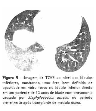
ResultadosOs achados mais comuns de TCAR em pacientes com pneumonia bacteriana pós-TMO foram os seguintes (Figuras 1 a 5): áreas de consolidação do espaço aéreo, em 18 (60%); pequenos nódulos centrolobulares, em 15 (50%); opacidade em vidro fosco, em 12 (40%); espessamento de parede brônquica, em 6 (20%); nódulos grandes, em 6 (20%); lesões pleurais, em 5 (16,7%); e opacidades em padrão de árvore em brotamento, em 3 (10%).


Dos 14 pacientes infectados com S. aureus, 7 (50%) apresentaram consolidação do espaço aéreo, 6 (42,9%) apresentaram pequenos nódulos centrolobulares, 4 (28,6%) apresentaram opacidade em vidro fosco, e 2 (14,3%) apresentaram opacidades em padrão de árvore em brotamento.
Entre os 9 pacientes infectados com P. aeruginosa, observou-se consolidação do espaço aéreo em 7 (77,8%), pequenos nódulos centrolobulares em 5 (55,6%), opacidade em vidro fosco em 3 (33,3%) e um grande nódulo escavado em 1 (11,1%).
Dos 7 pacientes restantes, 5 (71,4%) apresentaram opacidade em vidro fosco, 5 (71,4%) apresentaram espessamento de parede brônquica, 5 (71,4%) apresentaram nódulos grandes, 4 (57,4%) apresentaram consolidação do espaço aéreo, 4 (57,4%) apresentaram pequenos nódulos centrolobulares, e 1 (14,2%) apresentou opacidades em padrão de árvore em brotamento.
Os achados de TCAR nos pacientes com GVHD foram semelhantes àqueles obtidos na amostra como um todo.
As lesões pulmonares estavam distribuídas em áreas centrais e periféricas em 15 pacientes (50%), e estavam localizadas exclusivamente em áreas periféricas em 11 (36,7%). As lesões afetaram os lóbulos inferiores do pulmão em 22 casos (73,3%), os lóbulos médios em 20 (66,7%) e os lóbulos superiores em 5 (16,7%).
DiscussãoA infecção pulmonar é a causa mais comum de complicações após TMO. As complicações pulmonares pós-TMO seguem um cronograma relacionado ao estado imunológico dos pacientes. No período pré-enxerto (dias 0-30 após TMO), são comuns a neutropenia grave e as lesões da membrana da mucosa. Como resultado, infecções por vírus, fungos e bactérias, bem como por vírus respiratório sincicial, podem ocorrer durante esse período.(16,17) No período pós-enxerto (dias 31-100 após TMO), há imunidade humoral e celular comprometidas, sendo o citomegalovírus e o vírus respiratório sincicial os agentes causais mais comuns.(16,18) No período tardio pós-transplante (> 100 dias após o TMO), os pacientes geralmente apresentam função imune relativamente normal, e as infecções são mais frequentemente associadas à GVHD.
Nos pacientes com GVHD, os defeitos imunes podem persistir indefinidamente, com predisposição a infecções bacterianas, fúngicas e virais.(1,4,7,16-20) No presente estudo, a maioria (50%) dos pacientes apresentou pneumonia bacteriana no período tardio pós-transplante, em comparação a 26,7% no período pré-enxerto e 23,3% no período pós-enxerto.
Os organismos causadores de pneumonia bacteriana mais comuns, no período pré-enxerto após o TMO, são as bactérias gram-negativas da mucosa oral e intestinal, bem como organismos gram-positivos, especialmente Staphylococcus spp. e Streptococcus spp. No período pós-enxerto, as bactérias gram-negativas, incluindo P. aeruginosa e Escherichia coli, são as mais comuns, ao passo que as bactérias gram-positivas, tais como S. pneumoniae e S. aureus, são as mais comuns no período tardio pós-transplante.(6,7,10,16) Em nossa amostra de pacientes, a maioria dos casos deveram-se a S. aureus, P. aeruginosa ou S. viridans.
Fora realizados apenas alguns estudos objetivando avaliar achados de imagem em pacientes com pneumonia bacteriana pós-TMO. Os achados de radiografia de tórax assemelham-se àqueles vistos em pacientes imunocompetentes, com a predominância de consolidação do espaço aéreo.(6,10,16) Em um estudo de radiografias de tórax relacionadas a 52 episódios de pneumonia bacteriana pós-TMO,(20) observou-se infiltrado segmental em 37 (71%), infiltrado lobar em 10 (19%) e infiltrado em padrão patchy em 5 (10%). Em 12 episódios (23%), a infecção envolvia mais de um lóbulo ou segmento. Em 7 (13%), houve derrame pleural acompanhado de infiltrado do parênquima. Um grupo de autores estudou os achados de TCAR de 114 pacientes com pneumonia bacteriana, incluindo 35 pacientes imunodeprimidos.(14) As alterações mais comumente encontradas pelos autores foram a consolidação do espaço aéreo (em 85%), opacidade em vidro fosco (em 31%), opacidades reticulares (em 22%) e pequenos nódulos centrolobulares (em 17%).
Esses achados condizem com os obtidos no presente estudo, no qual 60% dos pacientes apresentaram consolidação do espaço aéreo e 40% apresentaram opacidade em vidro fosco. Entretanto, pequenos nódulos centrolobulares foram mais comuns em nosso estudo, sendo observados em 15 (50%) dos 30 casos. Na maioria de nossos pacientes, as alterações identificadas nas imagens de TCAR estavam localizadas nos lóbulos inferiores ou médios do pulmão. Observou-se derrame pleural em 10 pacientes, e nenhum dos pacientes apresentou linfonodomegalia. Não foram encontradas diferenças significativas entre as várias etiologias em termos de achados de TCAR.
Na amostra do nosso estudo, observamos que os achados de TCAR mais comuns em pacientes com pneumonia bacteriana pós-TMO foram a consolidação do espaço aéreo, pequenos nódulos centrolobulares e opacidade em vidro fosco, geralmente distribuídos nas regiões centrais e periféricas das zonas pulmonares médias e inferiores. Outros estudos são necessários, correlacionando tais achados com dados clínicos e laboratoriais, a fim de definir de modo mais completo o diagnóstico de pneumonia bacteriana após TMO, já que os achados de TCAR são semelhantes aos vistos em outras complicações pulmonares infecciosas após o procedimento.
Referências 1. Soubani AO, Miller KB, Hassoun PM. Pulmonary complications of bone marrow transplantation. Chest. 1996;109(4):1066-77.
2. Krowka MJ, Rosenow EC 3rd, Hoagland HC. Pulmonary complications of bone marrow transplantation. Chest. 1985;87(2):237-46.
3. Breuer R, Lossos IS, Berkman N, Or R. Pulmonary complications of bone marrow transplantation. Respir Med. 1993;87(8):571-9.
4. Wah TM, Moss HA, Robertson RJ, Barnard DL. Pulmonary complications following bone marrow transplantation. Br J Radiol. 2003;76(906):373-9.
5. Heussel CP, Kauczor HU, Heussel G, Fischer B, Mildenberger P, Thelen M. Early detection of pneumonia in febrile neutropenic patients: use of thin-section CT. AJR Am J Roentgenol. 1997;169(5):1347-53.
6. Escuissato DL, Gasparetto EL, Marchiori E, Rocha Gde M, Inoue C, Pasquini R, et al. Pulmonary infections after bone marrow transplantation: high-resolution CT findings in 111 patients. AJR Am J Roentgenol. 2005;185(3):608-15.
7. Winer-Muram HT, Gurney JW, Bozeman PM, Krance RA. Pulmonary complications after bone marrow transplantation. Radiol Clin North Am. 1996;34(1):97-117.
8. Worthy SA, Flint JD, Müller NL. Pulmonary complications after bone marrow transplantation: high-resolution CT and pathologic findings. Radiographics. 1997;17(6):1359-71.
9. Choi YH, Leung AN. Radiologic findings: pulmonary infections after bone marrow transplantation. J Thorac Imaging. 1999;14(3):201-6.
10. Gosselin MV, Adams RH. Pulmonary complications in bone marrow transplantation. J Thorac Imaging. 2002;17(2):132-44.
11. Mori M, Galvin JR, Barloon TJ, Gingrich RD, Stanford W. Fungal pulmonary infections after bone marrow transplantation: evaluation with radiography and CT. Radiology. 1991;178(3):721-6.
12. Gasparetto EL, Escuissato DL, Marchiori E, Ono S, Frare e Silva RL, Müller NL. High-resolution CT findings of respiratory syncytial virus pneumonia after bone marrow transplantation. AJR Am J Roentgenol. 2004;182(5):1133-7.
13. Gasparetto EL, Ono SE, Escuissato D, Marchiori E, Roldan L, Marques HL, et al. Cytomegalovirus pneumonia after bone marrow transplantation: high resolution CT findings. Br J Radiol. 2004;77(921):724-7.
14. Reittner P, Ward S, Heyneman L, Johkoh T, Müller NL. Pneumonia: high-resolution CT findings in 114 patients. Eur Radiol. 2003;13(3):515-21.
15. Hansell DM, Bankier AA, MacMahon H, McLoud TC, Müller NL, Remy J. Fleischner Society: glossary of terms for thoracic imaging. Radiology. 2008;246(3):697-722.
16. Leung AN, Gosselin MV, Napper CH, Braun SG, Hu WW, Wong RM, et al. Pulmonary infections after bone marrow transplantation: clinical and radiographic findings. Radiology. 1999;210(3):699-710.
17. Heussel CP, Kauczor HU, Heussel GE, Fischer B, Begrich M, Mildenberger P, et al. Pneumonia in febrile neutropenic patients and in bone marrow and blood stem-cell transplant recipients: use of high-resolution computed tomography. J Clin Oncol. 1999;17(3):796-805.
18. Chan CK, Hyland RH, Hutcheon MA. Pulmonary complications following bone marrow transplantation. Clin Chest Med. 1990;11(2):323-32.
19. Graham NJ, Müller NL, Miller RR, Shepherd JD. Intrathoracic complications following allogeneic bone marrow transplantation: CT findings. Radiology. 1991;181(1):153-6.
20. Lossos IS, Breuer R, Or R, Strauss N, Elishoov H, Naparstek E, et al. Bacterial pneumonia in recipients of bone marrow transplantation. A five-year prospective study. Transplantation. 1995;60(7):672-8.
Sobre os autoresLuiz Otávio de Mattos Coelho
Radiologista. Clínica Diagnóstico Avançado Por Imagem - DAPI - Curitiba (PR) Brasil.
Taísa Davaus Gasparetto
Residente de Radiologia. Universidade Federal Fluminense - UFF - Niterói (RJ) Brasil.
Dante Luiz Escuissato
Professor Adjunto. Universidade Federal do Paraná - UFPR - Curitiba (PR) Brasil.
Edson Marchiori
Professor Titular. Departamento de Radiologia, Universidade Federal Fluminense - UFF - Niterói (RJ) Brasil.
Trabalho realizado na Universidade Federal Fluminense - UFF - Niterói (RJ) Brasil e na Universidade Federal do Paraná - UFPR - Curitiba (PR) Brasil.
Endereço para correspondência: Edson Marchiori. Rua Thomaz Cameron, 438, Valparaíso, CEP 25685-120, Petrópolis, RJ, Brasil.
Tel 55 21 2629-9076. E-mail: edmarchiori@gmail.com
Apoio financeiro: Nenhum.
Recebido para publicação em 5/8/2008. Aprovado, após revisão, em 10/11/2008.


