ABSTRACT
Coccidioidomycosis, a fungal illness acquired by the inhalation of arthroconidia of Coccidioides sp., was first described in 1894. Coccidioidomycosis is mainly restricted to areas with arid climate, alkaline soil and low rainfall. Consequently, most of the reported cases in Brazil have occurred in the northeastern region. We report three cases of pulmonary coccidioidomycosis occurring between 2005 and 2006 in an endemic area in the state of Ceará, Brazil. The three patients were immunocompetent adult males, hunters of armadillos (Dasypus novemcinctus), with complaints of cough, fever, dyspnea and pleuritic pain. All three patients presented pulmonary involvement, and one also presented cutaneous lesions. Chest X-rays and CT scans of the patients revealed characteristic coccidioidomycosis lesions. The diagnosis was confirmed by serological testing. All of the patients evolved to cure after antifungal treatment.
Keywords:
Coccidioidomycosis; Lung diseases, fungal; Coccidioides.
RESUMO
A coccidioidomicose, uma doença fúngica adquirida através da inalação do agente Coccidioides sp. sob a forma de artroconídio, foi pela primeira vez descrita em 1894. Restringe-se principalmente a áreas de clima árido, solo alcalino e regiões de baixo índice pluviométrico. Não por acaso, a maioria dos casos descritos no Brasil ocorreu na região Nordeste. Relatam-se três casos de coccidioidomicose pulmonar ocorridos nos anos de 2005 e 2006, em zona endêmica no interior do Ceará. Todos eram homens imunocompetentes de idade adulta, adeptos à prática de caça a tatus (Dasypus novemcinctus) com queixas de tosse, febre, dispneia e dor pleurítica. Houve evoluções com comprometimento pulmonar e lesão cutânea foi observada em apenas um paciente. Todos apresentaram radiografia e TC de tórax com lesões características da coccidioidomicose. O diagnóstico foi confirmado através de teste sorológico. Todos evoluíram para cura após tratamento com antifúngico.
Palavras-chave:
Coccidioidomicose; Pneumopatias fúngicas; Coccidioides.
IntroductionCoccidioidomycosis is an illness caused by Coccidioides sp., a dimorphic fungus that lives in the soil and is found mainly in desert regions in the southeastern United States and northwestern Mexico.(1) Northeastern Brazil, due to its similar climate, is also an endemic region for coccidioidomycosis. To date, 24 cases have been reported in Brazil, and 12 of those cases occurred in the state of Ceará.
In nature, Coccidioides sp. is associated with semiarid environments (in which the dry season is quite long), high temperatures in the dry season and limited rainfall, concentrated in a short period of time. Therefore, coccidioidomycosis presents a limited geographic distribution, and transmission is restricted to a few months of the year.(2)
The fungus disseminates as arthroconidia and is inhaled together with soil dust. In Brazil, armadillo hunting is an activity that carries a significant risk for infection.
Pulmonary coccidioidomycosis is typically self-limiting but can evolve to chronicity and dissemination.(3) In a small number of individuals, the infection disseminates beyond the thoracic cavity, affecting the skin, bones, joints or soft tissues, and it can also result in chronic meningitis.(4)
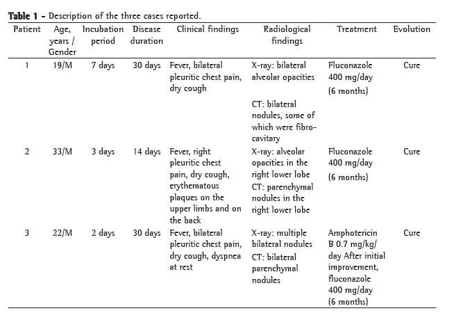
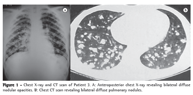
Case reportsThree male farmers, all mulattos and ranging from 19 to 33 years of age, who were armadillo hunters and residents of the northern region of the state of Ceará (Taperuaba-a district of the city of Sobral), were admitted to the Santa Casa de Misericórdia Hospital in the city of Sobral, Brazil, between December of 2005 and March of 2006. All three farmers had gone armadillo hunting every fifteen days in their region of residence. They presented the following symptoms: pleuritic pain; dry cough (two patients evolved to productive cough); fever; and dyspnea. Physical examination revealed rhonchi and crackles. Patient 1 had bilateral pulmonary involvement. Patient 2 developed cutaneous coccidioidomycosis, which manifested as diffuse erythematous plaques on the upper limbs. The same patient had pulmonary involvement (right lower lobe). Subsequently, the skin biopsy results confirmed the culture results (fungal growth on Sabouraud agar medium). Patient 3 developed more severe bilateral pulmonary involvement, presenting dyspnea at rest (SpO2, 85% on room air; Table 1). Chest X-rays and CT scans revealed diffuse bilateral opacities in Patients 1 and 3 (Figure 1). Bronchoscopy and bronchoalveolar lavage were then performed, and the samples were submitted to testing for acid-fast bacilli and neoplastic cells, both of which were negative. Due to epidemiological suspicion, a serological test (Ouchterlony double immunodiffusion), using positive control sera, was performed (Figure 2). The samples were collected one week after admission and analyzed at the Mycology Laboratory of the Medical Mycology Center of the Federal University of Ceará Department of Pathology and Legal Medicine. After the diagnosis was confirmed, treatment with an azole antifungal agent-fluconazole, 400 mg/day (6 months)-was initiated. The patient presenting the most severe condition was treated with amphotericin B, 0.7 mg/kg/day (total dose, 50 mg/day), combined with oxygen therapy. Subsequently, after clinical stabilization, fluconazole (at the same dose as that of the other two patients) was introduced. In the patient who developed cutaneous coccidioidomycosis, the erythematous plaques evolved to hyperchromic patches. After some weeks of treatment, the dermatological condition resolved. The treatment was successful for all three patients. Outpatient follow-up evaluations were performed at months 1, 2, 6 and 12 after discharge, and there was clinical and radiological improvement.


 Discussion
Although coccidioidomycosis was originally described in Argentina, its history is intimately associated with California, where a case was first described in 1894.(4) Until the end of the 1970s, Brazil was considered a coccidioidomycosis-free area. The first autochthonous cases in Brazil were reported in 1978 and 1979 in the states of Bahia and Piauí, respectively.(5,6) Approximately 15 years later, the first endemicity of this mycosis was described in Piauí.(7) Since then, the number of published case reports has increased considerably, and, in some of them, the association with armadillo hunting has also been described.(3,7,8)
Currently, coccidioidomycosis is considered endemic in the northeastern Brazilian states of Bahia, Piauí, Maranhão and Ceará.(9) In view of these case reports, it is imperative to consider this pathology in the differential diagnosis of insults with similar clinical profiles, such as TB, paracoccidioidomycosis, histoplasmosis and neoplasia.
Coccidioidomycosis is an endemic illness whose geographic distribution is relatively restricted to areas with arid or semiarid climate, typically alkaline soil (with high salinity) and low rainfall, which are conditions that foster the proliferation of its etiologic agent: Coccidioides sp.(4) Consequently, almost all cases of coccidioidomycosis reported in Brazil have occurred in the northeastern region.
The association between coccidioidomycosis and armadillo hunting has been described in the literature,(3,10) and the fungus has been isolated from armadillo tissue and from soil samples collected at armadillo burrows.(11) In northeastern Brazil, armadillos are used as food. When chased, these animals hide in their burrows, and hunters, who then dig the soil to capture them, are thereby susceptible to inhaling massive amounts of arthroconidia.(3)
Infection occurs after the inhalation of arthroconidia (infecting form of coccidioidomycosis), which, upon reaching the lungs, initiate their parasitic phase as thick-walled spherules containing endospores, each of which is able to form a new spherule, thereby resulting in exponential reproduction.(2)
Approximately 65% of the individuals who acquire this infection remain completely asymptomatic. Most of those who are symptomatic have pulmonary manifestations ranging from influenza-like diseases to severe pneumonia and septic syndrome. Although the most common manifestations are cough, fever, adynamia and chest pain, extrapulmonary forms with a miliary pattern, mainly affecting the skin, joints and meninges, can be seen.(12)
The radiological presentation ranges from alveolar or reticulonodular infiltrates, with or without pleural effusion, to multiple cavities, and there can be complications such as empyema and bronchopleural fistulae.(13)
The diagnosis is clinical, epidemiological and laboratorial. Laboratorial diagnosis is made based on the identification of the parasite in a direct mycological examination (sputum, pus, cerebrospinal fluid, bronchoalveolar lavage, skin lesion scraping and biopsy) or in a swab culture on Sabouraud agar medium.(4) Unfortunately, culture identification is slow and often impossible, since many patients with primary pulmonary infection are not able to expectorate secretion. Histopathological diagnosis can also be disadvantageous, since invasive sampling procedures can be dangerous. Therefore, the serologic tests developed by Smith (1948) are valuable in the diagnosis and follow-up treatment of patients with suspected coccidioidomycosis, particularly double immunodiffusion, which was established by Huppert & Bailey (1965) and is performed according to the Ouchterlony technique.(14) This technique is reliable and specific, as well as presenting few cross-reactions. It constitutes a qualitative test, since only an approximate quantification can be obtained by using serum dilutions in the study and by observing the maximum serum dilution forming precipitation bands against the antigen. This technique is less costly and more practical, as well as requiring only 24-72 h, thereby facilitating the institution of early and appropriate treatment.(10,15)
The use of antifungal therapy in acute infections (mild and moderate forms) is not mandatory, since, in most cases, the symptoms resolve spontaneously.(3,12,16) For those patients developing the pulmonary form or meeting the severity criteria, antifungal therapy, involving the use of oral azole antifungal agents (fluconazole or itraconazole) or amphotericin B (especially in cases of meningeal involvement), is recommended.(12,17) Although the appropriate duration of treatment remains controversial, reports of high recurrence rates after treatment discontinuation suggest that the medication should be maintained for six months or more.(3,12)
The comparison between the three cases reported here and the other cases reported in the literature revealed that the pattern of pulmonary manifestations was basically the same: diffuse pneumonia,(12) with different degrees of severity, and concomitant cutaneous dissemination, which is the most common extrathoracic form (one patient).(18) The radiological (X-ray and CT) findings were similar. Some invasive methods for making the diagnosis, such as lung biopsy(6) and lobectomy,(5) have been described in the literature. However, in the case of our patients, the use of such methods was not necessary, since the clinical profile, the imaging findings and the serology results were sufficient to confirm the diagnosis.
For our patients, due to the timing of the diagnosis, there was no need for treatments that were more invasive, and all three patients evolved to cure after treatment with antifungal therapy alone, unlike one published case, in which the fungus was detected by postmortem biopsy.(3)
The cases reported here alert us to the diagnostic possibility of coccidioidomycosis in patients with a history of exposure to soil in endemic areas and presenting radiological alterations consistent with coccidioidomycosis, together with respiratory symptoms. It can be expected that the dissemination of knowledge of the existence of this vast endemic area for coccidioidomycosis in northeastern Brazil will contribute to new cases being reported-cases that will certainly show the real significance of this mycosis in the regional nosology.
References 1. Martins Mdos A, de Araújo Eda M, Kuwakino MH, Heins-Vaccari EM, Del Negro GM, Vozza Júnior JA, et al. Coccidioidomycosis in Brazil. A case report. Rev Inst Med Trop Sao Paulo. 1997;39(5):299-304.
2. Moraes MA, Martins RL, Leal II, Rocha IS, Medeiros Jr P. Coccidioidomicose: novo caso brasileiro. Rev Soc Bras Med Trop. 1998;31(6):559-62.
3. Costa FA, Reis RC, Benevides F, Tomé GS, Holanda MA. Coccidioidomicose pulmonar em caçador de tatus. J. Pneumologia. 2001;27(5):275-8.
4. Ampel NM. Coccidioidomicose. In: Sarosi GA, Davies SF, editors. Doenças fúngicas do pulmão. Rio de Janeiro: Revinter; 2001. p. 57-76.
5. Gomes OM, Serrano RP, Prade HO, Barros Moraes NL, Varella AL, Fiorelli AI, et al. Coccidioidomicose pulmonar: primeiro caso nacional. Rev Assoc Med Bras. 1978;24(5):167-8.
6. Vianna H, Passos HV, Sant'ana AV. Coccidioidomicose: relato do primeiro caso ocorrido em nativo do Brasil. Rev Inst Med Trop São Paulo. 1979;21(1):51-5.
7. Wanke B. Coccidioidomicose. Rev Soc Bras Med Trop. 1994;27(Suppl 4):375-8.
8. Silva LC, Nunes LM, Sidrim JJ, Rios-Gonçalves AJ. Coccidioidomicose pulmonar aguda: primeiro surto epidêmico descrito no Ceará - segundo no Brasil. J Bras Med. 1997;72(5):49-66.
9. Wanke B, Lazera M, Monteiro PC, Lima FC, Leal MJ, Ferreira Filho PL, et al. Investigation of an outbreak of endemic coccidioidomycosis in Brazil's northeastern state of Piauí with a review of the occurrence and distribution of Coccidioides immitis in three other Brazilian states. Mycopathologia. 1999;148(2):57-67.
10. Veras KN, Figueiredo BC, Martins LM, Vasconcelos JT, Wanke B. Coccidioidomicose: causa rara de síndrome do desconforto respiratório agudo. J Pneumol. 2003;29(1):45-8.
11. Eulálio KD, de Macedo RL, Cavalcanti MA, Martins LM, Lazéra MS, Wanke B. Coccidioides immitis isolated from armadillos (Dasypus novemcinctus) in the state of Piauí, northeast Brazil. Mycopathologia. 2001;149(2):57-61.
12. Anstead GM, Graybill JR. Coccidioidomycosis. Infect Dis Clin North Am. 2006;20(3):621-43.
13. Stevens DA. Coccidioidomycosis. N Engl J Med. 1995;332(16):1077-82.
14. Camargo ZP. Técnicas imunológicas em Micologia. In: Sidrim JJ, Rocha MF, editors. Micologia médica à luz de autores contemporâneos. Rio de Janeiro: Guanabara Koogan; 2004. p. 349-357.
15. Iglesia de Elias Costa MR, Negroni R. Obtención de un nuevo antigeno del coccidioides immitis y desarrollo de un modelo experimental de enfermedad en ratas Wistar [Article in Spanish]. Rev Arg Mic. 1995;18(2):3-29.
16. Galgiani JN. Coccidioidomycosis: a regional disease of national importance. Rethinking approaches for control. Ann Intern Med. 1999;130(4 Pt 1):293-300.
17. Galgiani JN, Ampel NM, Catanzaro A, Johnson RH, Stevens DA, Williams PL. Practice guideline for the treatment of coccidioidomycosis. Infectious Diseases Society of America. Clin Infect Dis. 2000;30(4):658-61.
18. DiCaudo DJ. Coccidioidomycosis: a review and update. J Am Acad Dermatol. 2006;55(6):929-42; quiz 943-5.
Study carried out at the Sobral School of Medicine, Universidade Federal do Ceará - UFC, Federal University of Ceará - Sobral, Brazil.
Correspondence to: Fernando Moreira Batista Aguiar. Rua Oriano Mendes, 247, apto. 303, Centro, CEP 62010-370, Sobral, CE, Brasil.
Tel 55 88 3624-0315. E-mail: fernandombaguiar@hotmail.com
Financial support: None.
Submitted: 25 April 2008. Accepted, after review: 16 June 2008.
About the authorsRicardo Hideo Togashi
Professor in the Department of Pulmonology, Universidade Federal do Ceará - UFC, Federal University of Ceará - Sobral, Brazil.
Fernando Moreira Batista Aguiar
Medical Student. Sobral School of Medicine, Universidade Federal do Ceará - UFC, Federal University of Ceará - Sobral, Brazil.
Dalton Barros Ferreira
Medical Student. Sobral School of Medicine, Universidade Federal do Ceará - UFC, Federal University of Ceará - Sobral, Brazil.
Camille Matos de Moura
Medical Student. Sobral School of Medicine, Universidade Federal do Ceará - UFC, Federal University of Ceará - Sobral, Brazil.
Monique Teixeira Montezuma Sales
Medical Student. Sobral School of Medicine, Universidade Federal do Ceará - UFC, Federal University of Ceará - Sobral, Brazil.
Nikaelle Ximenes Rios
Medical Student. Sobral School of Medicine, Universidade Federal do Ceará - UFC, Federal University of Ceará - Sobral, Brazil.


