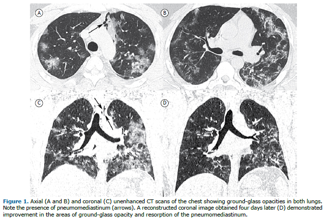
A 36-year-old male patient with no comorbidities was admitted to the ICU with a 14-day history of fever, cough, and intense dyspnea. On admission, his body temperature was 38°C (100.4°F), his RR was 30 breaths/min, and his SpO2 was 93%. Unenhanced CT of the chest showed predominantly peripheral areas of consolidation in both lungs, as well as pneumomediastinum (Figures 1A to 1C). Real-time fluorescence polymerase chain reaction of his sputum was positive for SARS-CoV-2 nucleic acid. After four days of hospitalization, having been treated exclusively with supportive measures, including oxygen therapy, the patient showed partial improvement of symptoms, and a new CT was performed (Figure 1D), which showed substantial diminution of the areas of consolidation and resorption of the pneumomediastinum.
The major causes of spontaneous pneumomediastinum include those related to the Valsalva maneuver and asthma. (1) The tomographic findings of COVID-19 have already been studied widely and reported in the medical literature.(2) To our knowledge, pneumomediastinum has been rarely associated with the disease.(3) As described in other diseases, the most probable mechanism of pneumomediastinum formation in the context of COVID-19 is the emergence of a pressure gradient between the alveoli and the surrounding structures, leading to alveolar rupture and air leak, which moves along the bronchovascular bundle until it reaches the mediastinum. This pressure gradient seems to be related to heterogeneous involvement of the lung when there are normal parenchymal areas adjacent to those affected by the disease.(1)
REFERENCES1. Araujo MS, Fernandes FL, Kay FU, Carvalho CR. Pneumomediastinum, subcutaneous emphysema, and pneumothorax after a pulmonary function testing in a patient with bleomycin-induced interstitial pneumonitis. J Bras Pneumol. 2013;39(5):613-619. https://doi.org/10.1590/S1806-37132013000500012
2. Simpson S, Kay FU, Abbara S, Bhalla S, Chung JH, Chung M, et al. Radiological Society of North America Expert Consensus Statement on Reporting Chest CT Findings Related to COVID-19. Endorsed by the Society of Thoracic Radiology, the American College of Radiology, and RSNA [published online ahead of print, 2020 Apr 28]. J Thorac Imaging. 2020;10.1097/RTI.0000000000000524. https://doi.org/10.1148/ryct.2020200152
3. Zhou C, Gao C, Xie Y, Xu M. COVID-19 with spontaneous pneumomediastinum. Lancet Infect Dis. 2020;20(4):510. https://doi.org/10.1016/S1473-3099(20)30156-0


 Read in Portuguese
 Portuguese PDF
 Print
Print
 Send this article by email
 How to cite this article
 Submit a comment
 Mendeley
 Pocket


