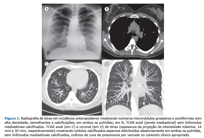
Uma restauradora de 29 anos de idade, que jamais fumara, apresentou ligeiro mal-estar e gripe durante alguns dias. A radiografia de tórax mostrou diversos nódulos de tamanhos variados, com alta densidade e distribuídos aleatoriamente em ambos os pulmões, semelhantes a calcificações (Figura 1A). Os resultados dos exames laboratoriais foram normais. Os testes de função pulmonar e a gasometria arterial foram normais: VEF1 = 3,23 l (97% do previsto); CVF = 3,81 l (98% do previsto); DLCO = 98%; pressão parcial de oxigênio = 97 mmHg; pressão parcial de dióxido de carbono = 39 mmHg. A TCAR de tórax realizada para esclarecer os achados da radiografia de tórax mostrou que, embora todos os nódulos estivessem calcificados, não foram encontrados linfonodos calcificados na TCAR axial (janela mediastinal, Figura 1B), e que havia numerosos nódulos bilaterais pequenos, bem definidos e distribuídos aleatoriamente em ambos os pulmões, sem espessamento intersticial ou qualquer outro achado patológico (Figuras 1C e 1D). Os resultados dos testes de função da paratireoide e de autoanticorpos foram negativos, assim como o foram os resultados do ensaio quantiFERON-TB (Cellestis, Ltd., Carnegie, Austrália); além disso, os níveis de cálcio no sangue estavam normais. A paciente confirmou uma infecção grave por varicela na infância (aos 4 anos de idade), e o teste de anticorpos para o vírus varicela-zóster apresentou resultados positivos (IgG = 348 mUI/ml e IgM = 0,34 mUI/ml). A varicela (catapora) é uma doença viral contagiosa transmitida por gotículas respiratórias. O surgimento de múltiplas calcificações nodulares pequenas e difusas em ambos os pulmões, com linfonodos não calcificados, é uma sequela incomum de pneumonia por varicela.
LEITURA RECOMENDADA1. Zanetti G, Hochhegger B, Marchiori E. Calcified multinodular lung lesions: a broad differential diagnosis. Clin Respir J. 2015 Jun 16. [Epub ahead of print] https://doi.org/10.1111/crj.12338
2. Marchiori E, Souza AS Jr, Franquet T, Müller NL. Diffuse high-attenuation pulmonary abnormalities: a pattern-oriented diagnostic approach on high-resolution CT. AJR Am J Roentgenol. 2005;184(1):273-82. https://doi.org/10.2214/ajr.184.1.01840273
3. Amin SB, Slater R, Mohammed TL. Pulmonary calcifications: a pictorial review and approach to formulating a differential diagnosis. Curr Probl Diagn Radiol. 2015;44(3):267-76. https://doi.org/10.1067/j.cpradiol.2014.12.005


 Read in English
 Portuguese PDF
 Print
Print
 Send this article by email
 How to cite this article
 Submit a comment
 Mendeley
 Pocket