To the Editor:The association of right heart thrombus with pulmonary thromboembolism (PTE) is uncommon, its incidence is underestimated, and treatment is still controversial. We report the case of a male patient with right heart thrombus and PTE.
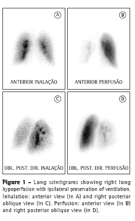
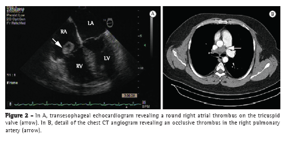
A 37-year-old man presented to the emergency room of the Hospital das Clínicas da Universidade Federal de Minas Gerais (HC-UFMG, Federal University of Minas Gerais Hospital das Clínicas), located in the city of Belo Horizonte, Brazil, with a two-day history of moderate dyspnea. Two years prior, the patient had had a similar episode, which was treated as bronchospasm. Six years prior, the patient had undergone left transtibial amputation because of popliteal artery occlusion. After one year of anticoagulation therapy (warfarin), he was diagnosed with muscular compression of the right popliteal artery, which was corrected surgically. At the time, factor VIII levels were found to be increased (200 IU/dL). The patient had a normal electrocardiogram. Physical examination revealed that the patient was alert, with an SpO2 of 90%, an HR of 72 bpm, and an arterial pressure of 150/100 mmHg. Cardiopulmonary examination was normal. A chest X-ray showed accentuation of the vascular markings on the left. A transthoracic echocardiogram (TTE) revealed a mobile thrombus (18 × 15 mm) attached to the posterior leaflet of the tricuspid valve and insinuating itself into the right ventricle (RV). Another thrombus (13 × 14 mm) was located in the inferior vena cava (IVC), diffuse right ventricular hypocontractility and impaired left ventricular diastolic relaxation having been observed. Lung scintigraphy (LS) revealed a homogeneous pattern of ventilation, markedly decreased right lung perfusion, and normal left lung perfusion (Figure 1). Although the patient had received anticoagulation therapy, he presented with persistent dyspnea and an SpO2 of 93%. A transesophageal echocardiogram revealed a large mobile thrombus located in the right atrium (RA) and projecting into the RV, as well as right ventricular dysfunction (Figure 2A). A chest CT angiogram revealed peripheral consolidations on the right and occlusion of the right pulmonary artery (2.9 cm), as well as filling defects in the upper lobe branch, middle lobe branch, lower lobe branch, segmental branch, and subsegmental branch, together with thrombi in six subsegmental branches of the left lower lobe (Figure 2B). A follow-up TTE, performed 17 days later, revealed an ejection fraction of 0.79, a dilated, hypokinetic RV, a pulmonary artery systolic pressure of 39 mmHg, and persistence of the thrombus in the RA. In that phase, oral anticoagulation therapy was maintained, in accordance with a decision made by the team of physicians, the patient, and his family. The patient was discharged on warfarin and was referred to the pulmonology department. He was readmitted in the following month because of worsening dyspnea. A second LS was performed, revealing a pattern that was similar to that which was revealed by the first. A TTE revealed that the intra-atrial thrombus and the thrombus in the IVC persisted, although they were smaller than the previous TTE had shown it to be. Because of those changes and symptoms, thrombolysis (alteplase, 100 mg in 2 h) was performed in the ICU. The procedure was uneventful. A transesophageal echocardiogram performed five days after the thrombolysis procedure showed that the thrombus in the RA had disappeared completely and that the thrombus (17 mm) in the IVC persisted. A follow-up LS performed 72 h later revealed no signs of lung reperfusion. His dyspnea having improved, the patient was discharged on warfarin and was referred to the HC-UFMG Department of Pulmonology for outpatient follow-up treatment.


Although mobile right heart thrombus is rarely identified in patients with PTE, its 3-23% incidence is probably underestimated because echocardiography is not systematically performed in those patients.(1,2) Because virtually 100% of mobile right heart thrombus cases are associated with PTE that is more severe, right heart thrombus in transit is a potential prognostic marker and should be differentiated from in situ right heart thrombus. The diagnosis is made by echocardiography. Right heart thrombi are classified into three types.
Type A thrombi are serpentine mobile thrombi associated with PTE/deep vein thrombosis (79-98%) and large vein thrombosis, migrating to the right heart. The clinical presentation is severe. The predisposing factors include tricuspid regurgitation, a prominent Eustachian valve, pulmonary hypertension, and low cardiac output.
Type B thrombi are immobile thrombi associated with an underlying heart disease. The association with PTE occurs in 38-40% of cases and has a less severe presentation.
Type C thrombi are extremely mobile non-serpentine thrombi that are similar to myxomas, as well as being intermediately and less strongly associated with PTE (62-67%).(3)
Type A thrombi are associated with high early mortality (42%); for type B thrombi, the mortality rate has been reported to be 4%.(3,4) Overall mortality is 31%, and it does not seem to be related to thrombus mobility but rather to the association of thrombus with severe PTE and to the type of treatment given. Therefore, the presence of mobile right heart thrombus might be a prognostic marker.(5)
There is still controversy regarding the best treatment option. The available data are from case series or consecutive case series, most of which suffer from a selection bias toward one of the forms of treatment. The treatment options include anticoagulation with heparin, use of thrombolytics, and surgical embolectomy.
In a systematic review involving 177 patients, the overall mortality rate was 27.1%, whereas the mortality rates for lack of treatment, use of anticoagulation therapy, surgical embolectomy, and treatment with thrombolytics were 100%, 28.6%, 23.8%, and 11.3%, respectively.(1) Treatment with thrombolytics exerted a protective effect when compared with anticoagulation with heparin (OR = 0.33; 95% CI: 0.11-0.98), as well as showing a trend toward a lower mortality rate (15.0%) than those for anticoagulation with heparin (37.5%) and embolectomy (29.0%), the same trend being observed when data from patients in consecutive case series were analyzed (use of thrombolytics vs. anticoagulation with heparin; p < 0.08).(1)
In another study, 42 (4%) of 1,113 patients with available echocardiographic data had cardiac thrombi and showed greater clinical impairment, whereas 1,071 had no cardiac thrombi and were considered controls. The overall 14-day mortality rates in the study and control groups were, respectively, 21% and 11% (p = 0.032), whereas the overall 3-month mortality rates were 29% and 16% (p = 0.036).(2) Although the mortality rates for the different types of treatment were similar in the study group (23.0%, 20.8%, and 25.0% for the patients treated with heparin, thrombolysis, and embolectomy, respectively), they were higher than were those observed in controls treated with heparin (mortality rate, 8%). This difference might be due to the fact that the patients with cardiac thrombus were more severely ill.(2)
In a prospective, consecutive case series of 335 patients with PTE admitted to the ICU, 12 (4%) had mobile right heart thrombus. Thrombolytics were administered to all but 3 patients, who died before infusion. In 7 of the 9 remaining patients, there were no signs of the thrombus or of right ventricular overload 12 h after infusion; 2 patients required an adjuvant procedure because the thrombus persisted. In all cases, there was improvement in lung perfusion, as revealed by LS and angiography, and this suggests that the use of thrombolytics (recombinant tissue plasminogen activator) produces a rapid effect in most patients.(6) In a series (n = 343) involving 18 patients (5.2%) with thrombus in transit, 16 were treated with thrombolytics, the thrombus having disappeared within less than 2 h after completion of the infusion, within 12 h after completion of the infusion, and within 24 h after completion of the infusion, respectively, in 50%, 25%, and 25% of the cases; therefore, because the response to thrombolytics is rapid in most cases, physicians should proceed with caution after the end of thrombolytic treatment.(7)
In the case described here, the fear of complications in a patient with severe vascular sequelae partially explains the conservative treatment initially given. However, thrombolysis, which had been indicated, proved effective in resolving the mobile thrombus; nevertheless, it did not lead to lung reperfusion, which should be achieved surgically. Therefore, because mobile right heart thrombus is associated with PTE that is more severe and because the mortality rates are high, thrombolytic treatment is indicated for selected patients with mobile right heart thrombus.
Thauana Luiza de Oliveira
Resident in Clinical Medicine,
Federal University of Minas Gerais Hospital das Clínicas,
Belo Horizonte, Brazil
Carolina Martins Vieira
Resident in Clinical Medicine,
Federal University of Minas Gerais Hospital das Clínicas,
Belo Horizonte, Brazil
Juliana Bernardes Costa
Resident in Pulmonology,
Federal University of Minas Gerais Hospital das Clínicas,
Belo Horizonte, Brazil
Tarciane Aline Prata
Resident in Pulmonology,
Federal University of Minas Gerais Hospital das Clínicas,
Belo Horizonte, Brazil
André Soares de Moura Costa
Medical Student,
Federal University of Minas Gerais School of Medicine,
Belo Horizonte, Brazil
Maria do Carmo Pereira Nunes
Professor,
Federal University of Minas Gerais School of Medicine,
Belo Horizonte, Brazil
Fernando Antônio Bottoni
Professor,
Federal University of Minas Gerais School of Medicine,
Belo Horizonte, Brazil
Ricardo de Amorim Corrêa
Professor,
Federal University of Minas Gerais School of Medicine, and
Head of the Department of Pulmonology and Thoracic Surgery, Federal University of Minas Gerais Hospital das Clínicas,
Belo Horizonte, BrazilReferences1. Rose PS, Punjabi NM, Pearse DB. Treatment of right heart thromboemboli. Chest. 2002;121(3):806-14. PMid:11888964. http://dx.doi.org/10.1378/chest.121.3.806
2. Torbicki A, Galié N, Covezzoli A, Rossi E, De Rosa M, Goldhaber SZ, et al. Right heart thrombi in pulmonary embolism: results from the International Cooperative Pulmonary Embolism Registry. J Am Coll Cardiol. 2003;41(12):2245-51. http://dx.doi.org/10.1016/S0735-1097(03)00479-0
3. The European Cooperative Study on the clinical significance of right heart thrombi. European Working Group on Echocardiography. Eur Heart J. 1989;10(12):1046-59. PMid:2606115.
4. Wood KE. Major pulmonary embolism: review of a pathophysiologic approach to the golden hour of hemodynamically significant pulmonary embolism. Chest. 2002;121(3):877-905. PMid:11888976. http://dx.doi.org/10.1378/chest.121.3.877
5. Kinney EL, Wright RJ. Efficacy of treatment of patients with echocardiographically detected right-sided heart thrombi: a meta-analysis. Am Heart J. 1989;118(3):569-73. http://dx.doi.org/10.1016/0002-8703(89)90274-3
6. Pierre-Justin G, Pierard LA. Management of mobile right heart thrombi: a prospective series. Int J Cardiol. 2005;99(3):381-8. PMid:15771917. http://dx.doi.org/10.1016/j.ijcard.2003.10.071
7. Ferrari E, Benhamou M, Berthier F, Baudouy M. Mobile thrombi of the right heart in pulmonary embolism: delayed disappearance after thrombolytic treatment. Chest. 2005;127(3):1051-3. PMid:15764793. http://dx.doi.org/10.1378/chest.127.3.1051


