To the Editor:Emphysematous bullae are defined as functionally inert, air-filled blebs that are larger than 1 cm and are covered by a thin outer layer that is either translucent or opalescent and is less than 1 mm thick, as well as consisting primarily of visceral pleura.(1) Under low pressure, lung bullae can either expand and lead to mediastinal compression or burst and cause hypertensive pneumothorax, massive hemorrhage, or gas embolism, characterizing an uncommon type of barotrauma.(2)
Cases of gas embolism in divers who ascend too rapidly from great depths constitute the most common example of barotrauma, cases of barotrauma during air travel being rare. Therefore, we report the case of a 62-year-old male patient who experienced acute respiratory failure due to expansion of a lung bulla during air travel.
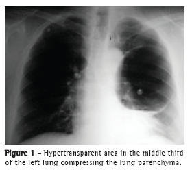
The patient was admitted to an emergency room with intense chest pain and sudden-onset dyspnea during air travel. Immediately after admission, electrocardiography was performed and cardiac enzyme levels were determined. The electrocardiogram and cardiac enzyme levels were found to be normal. A chest X-ray revealed a round hypertransparent area located in the middle third of the left lung and compressing the lung parenchyma (Figure 1). An axial CT scan of the chest confirmed the presence of a hypertransparent area occupying the entire anterior segment of the left upper lobe and measuring 12.2 × 9.0 cm, as well as containing a fine liquid layer. No other changes were found. The patient underwent left thoracotomy with selective lung intubation. We found a lung bulla located in the upper lobe and occupying the entire mediastinal surface of the lung. The affected area was resected and stapled with a mechanical stapler, and abrasion pleurodesis was performed. No other macroscopic changes were observed during the surgical procedure. After the surgical procedure, the patient was transferred to the ICU, where he was woken and extubated. The postoperative evolution was favorable, the chest tube having been removed on postoperative day 5. A follow-up chest X-ray revealed re-expansion of the left lung and hypotransparent areas in the middle third, which were attributed to the edema and the hematoma at the suture site. The histopathological features of the surgical specimen were found to be consistent with cystic adenomatoid malformation.
Cystic adenomatoid malformation of the lung is generally asymptomatic in adults, the diagnosis being incidental.(3) Changes in the atmospheric pressure can lead to rupture of previously asymptomatic lung bullae. This can be explained by Boyle's Law, according to which the volume of a gas is inversely proportional to its pressure.(4,5)

During commercial flights, the pressure in the passenger cabin varies according to the altitude. At an altitude of approximately 9,000 m, the airplane is pressurized in order to maintain an internal pressure of 2,400 m. During ascent, that change causes the pressure to drop from 760 mmHg (sea level) to 564 mmHg, a pressure that is reached within approximately 20 min after takeoff. The decrease in atmospheric pressure in that situation has two effects; it can cause expansion of the lung bulla leading to mediastinal compression, as was the case in our patient, or it can cause the pressure inside the bulla to rise above lung rupture pressure, which results in systemic embolism.(2) Although our patient was asymptomatic, he was aware of the lung bulla and was being followed periodically. However, he had never been alerted to the possibility of barotrauma.
Surgical treatment of lung bullae is indicated for patients who present with complications, such as rupture, pneumothorax, bleeding, and infection. In cases of uncomplicated lung bullae, resection is reserved for asymptomatic patients, for bullae occupying more than 30% of the hemithorax, and for progressively growing bullae.(6) Video-assisted thoracoscopic resection of lung bullae, when appropriate, is safe and effective.(7) One alternative that is less invasive and avoids complications related to general anesthesia is simple chest tube drainage under local anesthesia.(8) Regardless of the technique employed, surgical resection remains the treatment of choice for congenital cystic malformations of the lung and pulmonary lesions in which the radiological findings are inconclusive; the procedure allows us to conduct a histological study and prevent infections or neoplastic transformation.(9) In the case reported here, the patient presented with sudden-onset chest pain and dyspnea due to expansion of a lung bulla located in the upper lobe of the left lung, as confirmed by chest X-rays. According to the patient, previous imaging tests had shown the bulla to be smaller than we found it to be. Our choice of treatment was resection of the lung bulla by thoracotomy and mechanical stapling associated with abrasion pleurodesis.
Bullous lung disease has characteristics of benignity, and complications are uncommon. Variations in atmospheric pressure cause organic changes that can be more evident in pre-existing cavities, such as cystic cavities in the lung. Patients with lung bullae should be alerted to the possibility of expansion of the bullae in situations of marked variations in pressure, such as air travel and diving.
Fernando Luiz Westphal
Coordinator of the Teaching and Research Center,
Federal University of Amazonas
School of Medicine
Getúlio Vargas University Hospital, Manaus, Brazil
Luís Carlos de Lima
Chief Surgeon,
Department of Thoracic Surgery, Federal University of Amazonas
School of Medicine
Getúlio Vargas University Hospital, Manaus, Brazil
José Corrêa Lima Netto
Attending Physician,
Department of Thoracic Surgery, Federal University of Amazonas
School of Medicine
Getúlio Vargas University Hospital, Manaus, Brazil
Márcia dos Santos da Silva
Resident in Otolaryngology,
Federal University of Amazonas
School of Medicine,
Manaus, Brazil
Ingrid Loureiro de Queiroz Lima
Resident in Clinical Medicine,
Federal University of Amazonas
School of Medicine,
Manaus, Brazil
Danielle Cristine Westphal
Medical Student,
Federal University of Amazonas
School of Medicine,
Manaus, BrazilReferences1. Klingman RR, Angelillo VA, DeMeester TR. Cystic and bullous lung disease. Ann Thorac Surg. 1991;52(3):576-80. http://dx.doi.org/10.1016/0003-4975(91)90939-N
2. Neidhart P, Suter PM. Pulmonary bulla and sudden death in a young aeroplane passenger. Intensive Care Med. 1985;11(1):45-7. PMid:3968301. http://dx.doi.org/10.1007/BF00256066
3. Nai GA, Zelandi Filho C, Viero RM, Defaveri J. Malformação congênita adenomatóide cística do pulmão: relato de quatro casos. J Pneumol. 1998;24(5):335-8.
4. Belcher E, Lawson MH, Nicholson AG, Davison A, Goldstraw P. Congenital cystic adenomatoid malformation presenting as in-flight systemic air embolisation. Eur Respir J. 2007;30(4):801-4. PMid:17906087. http://dx.doi.org/10.1183/09031936.00153906
5. Mellem H, Emhjellen S, Horgen O. Pulmonary barotrauma and arterial gas embolism caused by an emphysematous bulla in a SCUBA diver. Aviat Space Environ Med. 1990;61(6):559-62. PMid:2369396.
6. Menconi GF, Melfi FM, Mussi A, Palla A, Ambrogi MC, Angeletti CA. Treatment by VATS of giant bullous emphysema: results. Eur J Cardiothorac Surg. 1998;13(1):66-70. http://dx.doi.org/10.1016/S1010-7940(97)00294-7
7. Lin KC, Luh SP. Video-assisted thoracoscopic surgery in the treatment of patients with bullous emphysema. Int J Gen Med. 2010;3:215-20. PMid:20830196. PMCid:2934603.
8. Saad Júnior R, Mansano MD, Botter M, Giannini JA, Dorgan Neto V. Tratamento operatório de bolhas no enfisema bolhoso: uma simples drenagem. J Pneumol. 2000;26(3):113-8. http://dx.doi.org/10.1590/S0102-35862000000300003
9. Morelli L, Piscioli I, Licci S, Donato S, Catalucci A, Del Nonno F. Pulmonary congenital cystic adenomatoid malformation, type I, presenting as a single cyst of the middle lobe in an adult: case report. Diagn Pathol. 2007;2:17. PMid:17555585. PMCid:1892770. http://dx.doi.org/10.1186/1746-1596-2-17


